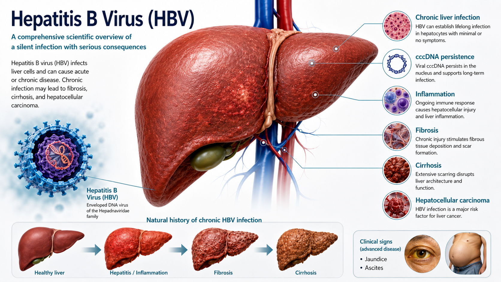
HEPATITIS B VIRUS (HBV)
Structured Abstract
Review Methodology and Evidence Framework
This publication is a structured narrative scientific review, not a systematic review, meta-analysis, or clinical practice guideline. The objective is to integrate established HBV biology with current epidemiology and selected rapidly evolving research areas while clearly separating well-established mechanisms from emerging or model-dependent findings.
At-a-Glance Molecular Snapshot
| Feature | Scientific summary |
|---|---|
| Taxonomy | Family Hepadnaviridae, genus Orthohepadnavirus; ICTV places human HBV in species Orthohepadnavirus hominoidei. |
| Virion | Enveloped Dane particle, typically about 40–45 nm; internal icosahedral capsid; three related envelope proteins (L, M, S). |
| Genome | approximately 3.2 kb relaxed circular, partially double-stranded DNA with extensively overlapping S, C, P, and X open reading frames. |
| Replication logic | Host RNA polymerase II transcribes cccDNA; pgRNA is encapsidated and reverse-transcribed to DNA inside nucleocapsids. |
| Entry | Initial attachment to heparan sulfate proteoglycans followed by high-specificity engagement of NTCP by myristoylated preS1. |
| Persistence | Nuclear cccDNA minichromosome plus long-lived infected-cell clones; integrated HBV DNA is a distinct genomic legacy. |
| Disease mechanism | Predominantly immune-mediated injury with contributions from inflammatory remodeling, fibrosis, viral proteins, integration, and epigenomic change. |
| Cancer link | HBV promotes HCC through chronic inflammation/regeneration plus direct genomic effects of integration and viral regulatory proteins. |
| Global burden | WHO 2026 estimates for 2024: approximately 240 million chronic infections, approximately 0.9 million new HBV infections, approximately 1.1 million HBV-related deaths. |
Molecular Lifecycle Roadmap
The HBV life cycle is cyclic rather than strictly linear, because newly synthesized viral genomes may either be secreted in virions or recycled to the nucleus to reinforce the cccDNA pool. The map below separates the major mechanistic checkpoints.
Detailed Contents
Hepatitis B virus (HBV) is one of the most consequential human viral pathogens. It is a highly infectious, hepatotropic DNA virus that can produce an acute infection or establish chronic infection lasting decades. Its long-term consequences include progressive fibrosis, cirrhosis, liver failure and hepatocellular carcinoma (HCC). The seriousness of HBV is amplified by a defining paradox: a person can carry a biologically active infection while appearing healthy and experiencing no obvious symptoms.
The absence of symptoms does not imply virological inactivity. Inside hepatocytes, HBV can establish covalently closed circular DNA (cccDNA) as a chromatinized nuclear reservoir, continue producing viral RNAs and proteins, generate new viral genomes through reverse transcription, and leave integrated HBV DNA fragments within the host genome. At the same time, immune responses can drive repeated cycles of hepatocyte injury and regeneration. These processes can unfold without jaundice, severe pain or other symptoms that would reliably warn a person that chronic liver disease is developing.
The 2026 World Health Organization (WHO) estimates place the burden at approximately a quarter of a billion people. WHO estimated that 240 million people were living with chronic hepatitis B in 2024, that about 0.9 million new HBV infections occurred that year, and that HBV caused approximately 1.1 million deaths—mostly from cirrhosis and HCC. That mortality corresponds to roughly 3,000 deaths every day. These are not remote historical numbers; they describe the current global burden.
A year-matched comparison with other major viral epidemics helps place this mortality burden in context. For a year-matched comparison, WHO estimated about 630,000 HIV-related deaths worldwide in 2024. In that same year, estimated HBV-related mortality was about 1.1 million—approximately 1.7 times as high. The diseases are biologically different and the comparison should not be used to diminish the importance of HIV; rather, it demonstrates how large the HBV burden is despite receiving far less public visibility in many settings. CDC also characterizes HBV as highly infectious and notes that infectious virus can persist on surfaces for at least seven days.
The burden is geographically unequal, and Ethiopia is part of the global high-burden picture. WHO reported in 2026 that ten countries—Bangladesh, China, Ethiopia, Ghana, India, Indonesia, Nigeria, the Philippines, South Africa and Viet Nam—accounted for 69% of HBV-related deaths worldwide in 2024. Ethiopia’s inclusion in this group should be understood as a warning that HBV is not a peripheral health issue: it is a major national and regional public-health challenge with global consequences.
Recent Ethiopian evidence reinforces that concern. A systematic review and meta-analysis published in 2025 included 72 studies with a combined 83,058 participants and estimated a pooled HBV prevalence of 6.9% (95% CI 6.1–7.7). As an intuitive illustration, 6.9% is close to seven people in every hundred. However, this pooled estimate must be interpreted scientifically: the included studies represented heterogeneous populations, regions and testing approaches, so it is not equivalent to a single nationally representative household survey and should not be applied mechanically to every community. Even with that caveat, the analysis documents a substantial and persistent HBV burden in Ethiopia.
Another reason for urgency is underdiagnosis. WHO estimated that only about 27% of people living with chronic HBV worldwide had been diagnosed by 2024. This creates a dangerous combination: a virus capable of long-term silent persistence, limited awareness of infection status, and liver disease that may become clinically obvious only after substantial fibrosis, cirrhosis or HCC has developed. The absence of symptoms therefore cannot be used as reassurance that infection or liver injury is absent.
Hepatitis B deserves coordinated attention across government, public-health institutions, hospitals, laboratories, clinicians, professional societies, universities, schools, media organizations, community leaders and families. The response must be scientifically accurate and sustained. Public communication should neither create panic nor normalize complacency. It should make clear that HBV is vaccine-preventable, can be detected through reliable testing, can be reduced through safer blood and injection practices, and can be interrupted through strong maternal, newborn and childhood prevention programs.
The purpose of this monograph is therefore larger than describing a virus. It connects the public-health emergency to the molecular biology that makes HBV so persistent: the 3.2-kb overlapping genome, the rcDNA-to-cccDNA transition, the HBx–DDB1–SMC5/6 axis, transcription and reverse transcription, capsid maturation, immune evasion and exhaustion, viral integration, clonal hepatocyte expansion, fibrosis, cirrhosis and hepatocarcinogenesis. Understanding HBV from molecule to population is essential because the same biological mechanisms that fascinate virologists are the mechanisms that ultimately shape preventable illness, cancer and death.
Hepatitis B virus (HBV) is an enveloped, hepatotropic DNA virus whose biology is unusually compact, efficient, and persistent. Its approximately 3.2-kilobase genome is among the smallest of human DNA viruses, yet it encodes a complete infection program through four extensively overlapping open reading frames. The virus reaches hepatocytes through receptor-guided entry, repairs its incoming relaxed circular DNA (rcDNA) into a nuclear covalently closed circular DNA (cccDNA) minichromosome, uses host RNA polymerase II to generate viral RNAs, and then copies an RNA intermediate back into DNA by reverse transcription inside newly assembled nucleocapsids. That combination—DNA genome, RNA intermediate, reverse transcription, and a stable nuclear episome—places HBV among the most biologically distinctive human pathogens.
HBV-associated liver injury is driven predominantly by virus-host immune interactions rather than by direct viral cytolysis alone. In many phases of infection, hepatocytes can contain abundant viral material without immediate cell death. Much of the liver injury arises from the host response to infected hepatocytes, especially virus-specific T-cell activity, inflammatory signaling, and repeated cycles of immune attack and tissue repair. At the same time, chronic antigen exposure can progressively weaken HBV-specific immune function through exhaustion, altered metabolism, inhibitory receptors, and an immunoregulatory liver environment. This tension between viral persistence and immune control explains why HBV may remain clinically silent for years while molecular and histological processes continue in the liver.
Two molecular features dominate long-term HBV persistence. The first is cccDNA, a chromatinized episomal DNA molecule in the hepatocyte nucleus that acts as the transcriptional reservoir for viral RNAs. The second is integration of fragments of HBV DNA into the human genome. Integration is not required for production of complete new HBV genomes and is usually a replication dead-end, but integrated sequences can remain biologically active, can produce viral proteins such as HBsAg or HBx-related products, can alter chromosome structure, and can contribute to hepatocellular carcinoma (HCC) through genomic instability, insertion near growth-regulatory genes, and interaction with chronic inflammation.
The global burden remains enormous. WHO estimated that 240 million people were living with chronic HBV infection in 2024, with approximately 0.9 million new infections and 1.1 million HBV-related deaths in that year, largely from cirrhosis and HCC. The Western Pacific and African regions carry particularly high burdens. The disease is therefore simultaneously a problem of molecular virology, immunology, hepatology, cancer biology, maternal-and-child health, laboratory medicine, and public health.
Modern HBV science emerged from a sequence of discoveries that connected epidemiology, serology, electron microscopy, enzymology, and molecular genetics. In 1965, the ‘Australia antigen’ was reported and was later recognized as hepatitis B surface antigen (HBsAg). This provided the first practical molecular handle on a disease that had long been known epidemiologically as serum-associated hepatitis.
In 1970, electron microscopy revealed the approximately 42-nm complete particle that became known as the Dane particle, alongside abundant smaller spherical and filamentous HBsAg-containing particles. The subsequent discovery of an endogenous DNA polymerase in HBV particles, cloning and sequencing of the genome, and recognition of reverse transcription transformed HBV from a serological phenomenon into a mechanistically defined virus.
A second wave of conceptual breakthroughs came from the identification of cccDNA as the persistent nuclear transcription template, the discovery of NTCP as a functional entry receptor in 2012, and the demonstration that HBx promotes cccDNA transcription by recruiting DDB1-dependent ubiquitin machinery to antagonize the SMC5/6 restriction complex. These discoveries explain, respectively, persistence, hepatocyte tropism, and a central mechanism of viral transcriptional control.
HBV remains a major global cause of chronic liver disease and primary liver cancer. WHO’s 28 July 2026 fact sheet estimates that 240 million people were living with chronic hepatitis B in 2024, with approximately 0.9 million new HBV infections and 1.1 million HBV-related deaths in that year. The Western Pacific and African regions carry the largest absolute burdens, with about 102 million and 64 million people living with chronic infection, respectively.
The age at acquisition strongly shapes the probability of persistence. WHO summarizes the contrast starkly: infection acquired in adulthood becomes chronic in fewer than 5% of cases, whereas infection in infancy and early childhood becomes chronic in about 95% of cases. This age dependence is one of the most important bridges between immunology and population epidemiology in HBV.
The burden is also hidden by underdiagnosis. WHO estimated that 65 million people—about 27% of those living with chronic HBV—were aware of their infection status in 2024. In the same year, global chronic HBV prevalence among children younger than five years was estimated at 0.6%, while the WHO African Region remained higher at about 1.4%.
The 2026 Global Hepatitis Report further emphasizes geographic inequality: the African Region accounted for 68% of new HBV infections in 2024, and birth-dose vaccination coverage in that region was only 17%. These figures explain why HBV remains simultaneously a virological, maternal-child-health, cancer-prevention, diagnostic, and health-systems challenge.
Africa carries a disproportionate share of global HBV transmission. WHO regional summaries place chronic HBV prevalence in the African Region at roughly 6% of the population, corresponding to about 65 million people in recent regional estimates, while the 2026 global fact sheet estimates about 64 million chronic infections for 2024. The difference reflects model updates and reporting time points rather than a biological contradiction.
A recent systematic review and meta-analysis of Ethiopian studies published from 2019 through early 2024 estimated a pooled HBV prevalence of 6.9% (95% CI 6.1–7.7). Such pooled estimates should be interpreted with care because underlying studies differ in region, study population, sampling frame, and assay strategy; they are best viewed as evidence of substantial national burden rather than as a single uniform prevalence for every community.
Molecular data from Ethiopia show important genetic diversity. A 2024 review reported genotype A as the most frequently described genotype, followed by genotype D; subgenotypes A1 and D2 were prominent, while D10 and less frequent genotypes or recombinants have also been reported. Because the number of whole-genome studies remains limited, national genotype maps are still incomplete and should not be overgeneralized from a small number of cohorts.
A major public-health milestone occurred in November 2025, when Ethiopia introduced a national hepatitis B birth-dose vaccine program. WHO reported an annual birth cohort of approximately 3.8 million newborns and an initial national coverage goal of at least 80%. This development is especially important because prevention of infection at birth interrupts the age window in which HBV is most likely to establish lifelong persistence.
For Ethiopian biomedical science, the next frontier is linkage: high-quality seroepidemiology, viral whole-genome sequencing, longitudinal liver phenotyping, maternal-infant cohorts, and standardized metadata should be analyzed together. This would allow local researchers to distinguish transmission history, genotype structure, immune phenotype, liver outcomes, and geographic heterogeneity rather than importing conclusions from unrelated populations.
HBV belongs to the family Hepadnaviridae and the genus Orthohepadnavirus. In current ICTV taxonomy, human HBV is placed within the species Orthohepadnavirus hominoidei. Hepadnaviruses are assigned to the realm Riboviria because their polymerases are evolutionarily related to reverse-transcribing enzymes, even though mature HBV particles carry DNA rather than RNA as the genome. Members of Hepadnaviridae share a compact partially double-stranded DNA genome and an obligatory reverse-transcription step in replication.
Orthohepadnaviruses infect mammals and typically have narrow host ranges. Natural HBV infection is associated with humans and certain non-human hominoids. This narrow tropism reflects several barriers, including receptor compatibility, intracellular host factors, innate restriction systems, and species-specific interactions required for viral transcription and replication. The discovery of the hepatocyte receptor sodium taurocholate cotransporting polypeptide (NTCP) was especially important because it explained a major component of liver tropism and species restriction.
HBV evolution is unusually dynamic for a DNA virus because replication proceeds through an error-prone reverse transcriptase that lacks the high-fidelity proofreading typical of many cellular DNA polymerases. As a result, every chronically infected person can contain a swarm of closely related viral variants—a quasispecies—rather than a single perfectly uniform sequence. Selection by host immunity, transmission bottlenecks, cellular environment, and stochastic processes continually reshapes this population.
Evolutionary ecology and species tropism
HBV belongs to a broader evolutionary landscape of mammalian and avian hepadnaviruses. Related orthohepadnaviruses occur in primates, bats, rodents, and other mammals, but species barriers are shaped by receptor compatibility, hepatocyte differentiation state, intracellular host factors, and immune restriction. The existence of related hepadnaviruses is scientifically important for reconstructing viral evolution and for model development; it does not imply that ordinary contact with those animals is a recognized route of human HBV transmission.
The human HBV genome has also been shaped by long-term geographic diversification, recombination, founder effects, and host population history. Phylogenetic classification is therefore a model of evolutionary relatedness, not a simple label for disease severity. Genotype, subgenotype, recombinant structure, within-host quasispecies, and specific mutations represent different levels of variation and should not be collapsed into a single category.
The complete infectious HBV virion, historically called the Dane particle, is an enveloped particle approximately 40–45 nm in diameter. Beneath its host-derived lipid envelope lies an icosahedral nucleocapsid built mainly from hepatitis B core protein (HBc). Inside the capsid are the viral DNA genome and the viral polymerase. The external envelope contains three related surface proteins—large (L), middle (M), and small (S) hepatitis B surface proteins—produced from the same surface open reading frame through alternative start sites.
A striking feature of HBV biology is that infected hepatocytes release huge numbers of non-infectious subviral particles (SVPs) containing surface antigen but lacking a nucleocapsid and viral genome. These particles occur as small spheres and as filaments. They can exceed complete virions by several orders of magnitude in blood. SVPs dominate the circulating HBsAg pool and contribute substantially to the antigenic environment encountered by the immune system; they also demonstrate the large biosynthetic investment HBV makes in non-infectious envelope particles. Modern structural studies using cryo-electron microscopy continue to reveal the organization of lipids and surface proteins within these particles.
The three envelope proteins have different domain architectures. The S protein forms the major mass of the envelope and SVPs. The M protein contains the S domain plus the preS2 region. The L protein contains preS1, preS2, and S; its N-terminal preS1 region is critical for high-affinity interaction with NTCP during entry. The relative orientation of L protein domains can change during assembly and infection, allowing the same molecule to contribute to both virion morphogenesis and receptor engagement.
The capsid can assemble in more than one icosahedral geometry. T=4 capsids containing 240 HBc monomers are common, while T=3 particles containing 180 monomers also occur. Core-protein structural plasticity has functional consequences: phosphorylation, nucleic-acid content, polymerase/pgRNA loading, and maturation state influence capsid behavior across the replication cycle.
HBV-infected cells release more than one extracellular particle class. In addition to genome-containing virions and classic HBsAg-rich subviral spheres and filaments, studies describe empty virions, naked capsids in some experimental or biological contexts, and RNA-containing particles. These populations complicate the simple assumption that every circulating viral antigen or particle directly represents an infectious genome-containing virion.
| Extracellular form | Contains envelope? | Contains capsid? | Typical nucleic acid / meaning |
|---|---|---|---|
| Dane virion | Yes | Yes | Predominantly rcDNA; infectious complete particle. |
| Spherical SVP | Yes (HBs proteins) | No | No viral genome; major source of circulating HBsAg. |
| Filamentous SVP | Yes (HBs proteins) | No | No viral genome; enriched in larger surface proteins relative to many spheres. |
| Empty virion | Yes | Yes | Capsid with little or no mature viral DNA; illustrates imperfect coupling of morphogenesis and genome completion. |
| RNA-containing particle | Usually enveloped particle forms described | Yes/variable | Can carry HBV RNA species, including pgRNA-related molecules; biological composition is heterogeneous. |
The HBV genome is a relaxed circular DNA molecule of roughly 3.2 kb. The minus strand is essentially genome length, whereas the plus strand is incomplete and variable in length. Viral polymerase is covalently attached to the 5′ end of the minus strand, and a short RNA primer remains associated with the 5′ end of the plus strand. This chemically unfinished genome is not immediately equivalent to a cellular plasmid; after nuclear delivery it must be repaired into cccDNA.
The genome contains four major, extensively overlapping open reading frames: S, C, P, and X. The S region encodes the L, M, and S surface proteins. The C region encodes the core protein and, from an upstream precore start, the precursor of HBeAg. The P region encodes the multifunctional polymerase, including terminal-protein, spacer, reverse-transcriptase, and RNase H domains. The X region encodes HBx, a small regulatory protein essential for efficient transcription from cccDNA in authentic infection systems. Because these reading frames overlap, a single nucleotide substitution can affect more than one viral protein or regulatory element, creating unusual evolutionary constraints.
HBV gene expression is controlled by multiple promoters and two major enhancers embedded within the compact genome. Host transcription factors that are enriched in hepatocytes help confer tissue specificity. The virus therefore achieves a high information density: protein-coding sequences, promoters, enhancers, direct repeats required for reverse transcription, encapsidation signals, and regulatory elements are interleaved across only a few thousand nucleotides.
The compact genome is organized around a highly constrained principle: coding regions, promoters, enhancers, regulatory motifs, and replication signals overlap physically. Consequently, a nucleotide change can influence several layers at once—for example, a polymerase amino-acid change may simultaneously alter the overlapping surface-protein sequence, and a promoter-region change may modify both transcription and a protein-coding region.
Regulatory architecture: promoters, enhancers, and RNA signals
HBV transcription is coordinated by several cis-regulatory elements embedded inside the coding genome. Major promoter systems include the core promoter, preS1 promoter, preS2/S promoter, and X promoter, while enhancer I and enhancer II increase transcription in a strongly hepatocyte-dependent regulatory environment. Because these elements overlap coding sequences and each other functionally, mutations can alter gene expression without changing the gross organization of the genome.
The genome also encodes RNA-level and replication signals. The epsilon stem-loop near the 5-prime end of pgRNA is recognized by viral polymerase and is essential for selective packaging and protein priming. Direct repeats DR1 and DR2 help organize template switches during DNA synthesis. HBV transcripts share a common 3-prime region and polyadenylation logic, and the post-transcriptional regulatory element (PRE), present in viral RNAs, contributes to RNA stability, processing, and export. These features explain how a genome of only about 3.2 kb controls a disproportionately complex RNA and replication program.
| Regulatory element | Primary molecular role |
|---|---|
| Core promoter / enhancer II | Controls precore and pgRNA transcription; closely linked to replication phenotype and HBeAg biology. |
| Enhancer I / X promoter region | Supports liver-enriched transcriptional control and X-gene expression. |
| preS1 and preS2/S promoters | Control the relative production of large, middle, and small surface-protein transcripts. |
| Epsilon (ε) RNA signal | Polymerase recognition, pgRNA packaging, and initiation of protein-primed reverse transcription. |
| DR1 / DR2 | Direct-repeat signals used during template switching and circularization steps of DNA synthesis. |
| PRE | Post-transcriptional regulation of viral RNA stability, export, and expression. |
HBV proteins are multifunctional and tightly coordinated. Surface proteins build the viral envelope, mediate entry, and form SVPs. HBc self-assembles into capsids, binds nucleic acids, participates in intracellular trafficking, and interfaces with host proteins. HBeAg is derived from the precore pathway and is secreted; it is not a structural component required to build the virion, but it is strongly associated with the immunological ecology of infection. Polymerase executes protein priming, reverse transcription, and RNase H activity. HBx acts as a transcriptional enabler by counteracting host restriction of cccDNA and also influences diverse cellular pathways relevant to persistence and carcinogenesis.
| Protein / antigen | Genomic origin | Core biological role |
|---|---|---|
| LHBs | preS1 + preS2 + S | Envelope protein; preS1 carries the NTCP-binding determinant; required for infectivity and participates in virion assembly. |
| MHBs | preS2 + S | Envelope/SVP component; contributes to envelope architecture and secretion. |
| SHBs | S | Major HBsAg component; abundant in virions and SVPs; principal antigenic basis of recombinant vaccines. |
| HBc | C ORF | Capsid assembly, pgRNA/polymerase packaging, nucleic-acid organization, intracellular trafficking, and genome replication compartment. |
| HBeAg | Precore pathway | Secreted core-related antigen; associated with replicative state but not a perfect proxy for HBV DNA; has immunological effects. |
| Polymerase | P ORF | Terminal-protein priming, reverse transcriptase, RNase H, epsilon recognition, and DNA synthesis. |
| HBx | X ORF | Supports transcription from cccDNA, prominently through DDB1-dependent antagonism of SMC5/6; has broader host-regulatory effects. |
HBc and HBeAg arise from the same precore/core genomic region but are biologically distinct products. Translation beginning at the core start codon produces HBc, which remains intracellular and assembles into capsids. Translation of the upstream precore sequence produces a precursor with an N-terminal signal peptide that enters the secretory pathway, is proteolytically processed, and is released as HBeAg.
Because HBeAg is secreted rather than incorporated as the structural core of the mature virion, HBeAg positivity should not be equated mechanically with the presence of a capsid protein in blood. HBeAg often accompanies high replication, but precore stop-codon variants and basal-core-promoter variants can markedly reduce or abolish HBeAg expression while viral replication continues.
HBeAg also has immunological significance. Experimental and clinical literature supports immunomodulatory and tolerogenic associations, particularly in early-life infection. These effects are one reason the precore pathway is discussed not only as a diagnostic marker system but also as part of the virus-host dialogue that influences chronicity.
HBV entry is a multistep process rather than a single receptor-binding event. Initial low-affinity attachment involves heparan sulfate proteoglycans on the hepatocyte surface. The virus then engages NTCP, a bile-acid transporter expressed on differentiated hepatocytes, through the myristoylated preS1 region of the large surface protein. NTCP binding is a major determinant of hepatotropism and species specificity. Additional host factors, including epidermal growth factor receptor (EGFR)-linked pathways, have been implicated in organizing receptor complexes and internalization.
After receptor engagement, the virion is internalized through endocytic pathways. Membrane fusion and uncoating release the nucleocapsid into the cytoplasm. HBV entry depends on coordinated receptor binding, endocytosis, and hepatocyte membrane trafficking that deliver the nucleocapsid from the extracellular compartment toward the nuclear pore.
The identification of NTCP transformed experimental HBV research because hepatoma-derived cell lines that express NTCP can support authentic entry much more effectively than earlier systems. However, receptor expression alone does not reproduce every aspect of infection; hepatocyte differentiation state, polarity, innate signaling, and other host factors remain important. This is one reason primary human hepatocytes, stem-cell-derived hepatocyte-like systems, organoids, and humanized animal models remain central to HBV research.
NTCP is physiologically a bile-acid transporter concentrated on the basolateral membrane of differentiated hepatocytes. Its expression state helps explain why many transformed hepatoma lines are poorly susceptible unless NTCP is restored and why differentiated hepatocyte identity is central to authentic HBV infection models. Receptor engagement is necessary but not sufficient for the entire entry program; membrane trafficking, endosomal routing, envelope fusion events, and capsid release remain active areas of investigation.
Once the viral envelope is removed, the nucleocapsid must move through the cytoplasm and deliver rcDNA to the nucleus. Capsid phosphorylation state, microtubule-associated transport, interactions with nuclear-pore components, and capsid maturation influence this process. Mature capsids carrying rcDNA are competent for nuclear delivery, whereas immature capsids containing RNA or incomplete replication products are handled differently.
At or near the nuclear pore, capsids undergo structural transitions that permit release of rcDNA into the nucleus. The exact timing and physical mechanism of uncoating remain active research areas. Importantly, nuclear arrival is not the end of the entry process: the incoming rcDNA carries several lesions and covalent attachments that must be repaired before it becomes the stable transcriptional template characteristic of HBV persistence.
A key conceptual uncertainty is whether mature capsids disassemble in a single defined step or undergo progressive structural remodeling at the nuclear pore complex. Core-protein phosphorylation, genome maturation state, capsid stability, and interaction with nuclear transport machinery all appear to influence genome delivery. The field therefore increasingly treats ‘uncoating’ as a regulated transition rather than as a simple rupture event.
Formation of covalently closed circular DNA is one of the most important events in HBV biology. Incoming rcDNA contains a complete minus strand linked to viral polymerase, an incomplete plus strand, an RNA primer, and discontinuities in both DNA strands. Host DNA-repair activities remove the attached polymerase and RNA primer, complete the plus strand, process abnormal ends, and ligate both strands into a supercoiled, covalently closed circle.
The resulting cccDNA is not integrated into the host chromosome. It persists as an episome in the hepatocyte nucleus and associates with histones and other chromatin proteins to form a minichromosome. This chromatinized organization allows cccDNA transcription to respond to epigenetic marks, host transcription factors, viral proteins, inflammatory signaling, and the metabolic state of the hepatocyte.
cccDNA is central to persistence because it can remain transcriptionally active or relatively silent for long periods and can be replenished when newly synthesized rcDNA-containing capsids recycle back to the nucleus. Its copy number is usually low on a per-cell basis, but even a small reservoir distributed across many hepatocytes can sustain long-lasting viral potential. The stability of cccDNA is influenced by hepatocyte death, cell division, immune pressure, and intracellular turnover, and its exact longevity in human liver remains an area of intense investigation.
Recent work has sharpened the view of cccDNA biogenesis as a host DNA-repair problem. Incoming rcDNA must lose the covalently attached viral polymerase from the minus strand, remove an RNA primer remnant from the plus strand, eliminate terminal redundancies or flap structures, complete the incomplete plus strand, and seal remaining nicks. Multiple host factors have been implicated, including TDP2 and FEN1 in selected processing steps and several DNA polymerases, PCNA/RFC-associated machinery, and DNA ligases. The relative contribution of individual factors can vary by experimental system, and no single enzyme should be treated as the sole universal ‘cccDNA enzyme.’
cccDNA behaves as a chromatin template. It associates with canonical histones and can acquire histone acetylation, methylation, and other epigenetic marks that correlate with transcriptional activity. Host transcription factors such as HNF family members, nuclear receptors, and other liver-enriched regulators act on HBV promoters and enhancers. The cccDNA minichromosome is therefore embedded within the same general regulatory logic used by the hepatocyte to control cellular genes, even though the viral DNA remains physically separate from host chromosomes.
A major host restriction factor is the structural maintenance of chromosomes 5/6 complex (SMC5/6), which can associate with episomal DNA and repress HBV transcription. HBx counteracts this restriction by binding DDB1 within a CUL4-containing ubiquitin-ligase system and redirecting it toward SMC5/6, leading to SMC5/6 degradation and relief of cccDNA silencing. This HBx–DDB1–SMC5/6 axis is now regarded as a central molecular mechanism enabling robust HBV gene expression.
The HBx story also illustrates a deeper principle: successful HBV persistence depends on precise manipulation of host nuclear quality-control systems. HBx is only about 17 kDa, yet its effect on cccDNA transcription is disproportionately large. Beyond SMC5/6, HBx has been linked to transcriptional regulation, DNA-damage responses, mitochondrial signaling, cell-survival pathways, and epigenetic changes. Not every reported HBx interaction is equally established in vivo, so mechanistic interpretation requires careful distinction between reproducible core functions and context-dependent effects observed in experimental models.
The term ‘minichromosome’ is literal at the molecular level: cccDNA associates with histones and non-histone proteins, acquires activating or repressive histone marks, and is influenced by chromatin remodelers, transcription factors, DNA methylation state, non-coding RNAs, and RNA modifications. This helps explain why identical amounts of intrahepatic cccDNA need not produce identical amounts of viral RNA.
cccDNA quantity and cccDNA activity are therefore distinct variables. A hepatocyte may contain cccDNA that is highly transcriptionally active, partially repressed, or deeply silenced. Conversely, HBsAg can also arise from integrated HBV DNA, so serum HBsAg concentration cannot be interpreted as a pure one-to-one measurement of cccDNA copy number.
One of the most important mechanistic advances in HBV biology was the identification of SMC5/6 as a host restriction complex that can repress transcription from extrachromosomal DNA, including HBV cccDNA. HBx counteracts this restriction by binding the cellular DDB1-containing ubiquitin-ligase machinery and redirecting it toward SMC5/6, promoting loss of the restriction complex and enabling robust viral transcription.
This mechanism explains why HBx is disproportionately important despite its small size. HBx does not function as a classical structural protein; rather, it changes the nuclear regulatory environment so that the viral episome can be expressed. The model also clarifies why HBx-deficient HBV can be profoundly impaired after authentic infection even when some replication steps can be reproduced in artificial transfection systems.
The HBx–SMC5/6 interaction should not be reduced to a single binary switch. HBx has additional reported interactions involving transcriptional control, DNA-damage responses, signaling, mitochondrial biology, and epigenetic regulation. Some of these effects may be context-dependent, and distinguishing physiologically relevant HBx functions from overexpression artifacts remains an important research challenge.
The canonical replication-competent HBV RNA program is transcribed from cccDNA by host RNA polymerase II. The principal cccDNA-derived transcripts include two closely related 3.5-kb RNAs—the precore RNA and pregenomic RNA (pgRNA)—plus approximately 2.4-kb and 2.1-kb surface transcripts and an approximately 0.7-kb X transcript. These RNAs share common 3′-end processing features but differ in their 5′ origins, coding capacity, and roles in the viral lifecycle.
The pgRNA is exceptional because it serves both as an mRNA for core protein and polymerase and as the RNA template that is packaged into nucleocapsids and reverse-transcribed into viral DNA. This dual role links nuclear transcription directly to cytoplasmic genome replication.
Integrated HBV DNA can independently generate selected viral transcripts, particularly surface-antigen-related RNAs and some HBx-related transcripts. Integrated sequences are generally defective for the complete canonical replication cycle, but their transcriptional activity can sustain HBsAg production and selected viral gene products independently of active cccDNA-driven genome replication. This distinction is especially important in long-standing chronic infection and when interpreting HBsAg as a biomarker of residual viral activity. [14]
HBV RNAs are further shaped by co-transcriptional and post-transcriptional processes including RNA splicing, polyadenylation, nuclear export, RNA stability, RNA-binding proteins, translation control, and chemical modifications such as N6-methyladenosine. These regulatory layers increase transcriptomic complexity without increasing genome size. [9,31,32]
HBV transcripts are more diverse than the canonical 3.5-kb, 2.4-kb, 2.1-kb, and 0.7-kb classes suggest. A proportion of HBV RNAs undergo alternative splicing, generating multiple spliced RNA species. Some can be packaged and reverse-transcribed into defective or rearranged DNA products, while others may encode noncanonical proteins such as the hepatitis B splice-generated protein described in experimental literature.
Post-transcriptional regulation includes RNA stability, nuclear export, ribosome access, RNA-binding proteins, microRNA interactions, and chemical modifications including N6-methyladenosine (m6A). These layers can change RNA half-life, translation efficiency, and replication competence without altering the underlying cccDNA sequence.
Spliced HBV RNAs are generally minor compared with canonical transcripts, and their abundance varies by viral genotype, cell state, and disease context. These transcripts demonstrate that the HBV transcriptome comprises a regulated population of RNA isoforms rather than a fixed set of full-length RNAs.
In the cytoplasm, pgRNA is translated to produce core protein (HBc) and viral polymerase. Selective packaging begins when polymerase recognizes the epsilon (ε) stem-loop near the 5′ end of pgRNA. Polymerase binding, together with host chaperone-assisted conformational changes, distinguishes pgRNA destined for replication from the larger pool of viral RNAs used only for translation.
Core dimers assemble around the polymerase–pgRNA complex to form the nucleocapsid. Reverse transcription is initiated by protein priming: a tyrosine residue in the terminal-protein domain of polymerase becomes covalently linked to the first nucleotide of nascent minus-strand DNA. The subsequent DNA-synthesis steps are described in Section 19 to avoid duplicating the reverse-transcription pathway here.
HBc is a dynamic structural and regulatory protein. Its N-terminal assembly domain forms dimers that build the icosahedral shell, whereas the arginine-rich C-terminal domain binds nucleic acids and is regulated by phosphorylation. Most capsids adopt T=4 symmetry with 240 HBc monomers, while T=3 particles containing 180 monomers also occur.
Core-protein phosphorylation and dephosphorylation influence pgRNA packaging, reverse transcription, intracellular trafficking, and nuclear events. The capsid is therefore not a passive container; it is a regulated reaction chamber whose structural state changes as the viral genome matures.
Capsid assembly must balance speed, selectivity, and stability. Empty capsids and other noncanonical particles can form, illustrating that assembly and genome packaging are tightly coordinated but not absolutely coupled.
Reverse transcription occurs within the nucleocapsid after protein priming has begun. The short nascent DNA initiated on epsilon is transferred to the 3′ direct-repeat region of pgRNA, after which minus-strand DNA elongation proceeds. The polymerase RNase H domain degrades most of the RNA template during minus-strand synthesis while preserving a short RNA oligomer that later serves as the primer for plus-strand DNA synthesis.
Additional template switches involving the direct-repeat sequences DR1 and DR2 position the plus-strand primer and establish the circular geometry of the genome. Plus-strand synthesis is usually incomplete when the virion is secreted, producing the characteristic relaxed circular DNA (rcDNA) genome. Alternative primer translocation can instead generate double-stranded linear DNA (dslDNA), which has important consequences for integration.
HBV reverse transcriptase lacks the high-fidelity proofreading typical of many cellular DNA polymerases. Replication therefore generates sequence variation that contributes to within-host quasispecies, genotype diversification, immune selection, and the emergence of biologically important variants.
Most mature HBV genomes adopt the relaxed circular DNA (rcDNA) configuration, but a minority follow an alternative priming pathway that produces double-stranded linear DNA (dslDNA). dslDNA is biologically important because it is a major precursor for HBV DNA integration into host chromosomes, linking reverse-transcription mechanics to long-term genomic consequences.
rcDNA and dslDNA can both participate in nuclear events, but their typical outcomes differ. rcDNA is optimized for repair into cccDNA, whereas dslDNA more readily enters host DNA-repair pathways that can join viral ends to chromosome breaks. Some dslDNA can also be circularized, but the resulting forms may be structurally different from canonical cccDNA.
This branch point explains an important principle: HBV persistence has two molecular legacies generated from related replication intermediates. cccDNA preserves the episomal, replication-competent transcription template, whereas integration preserves selected viral DNA fragments within the cellular genome and can shape antigen production, clonal selection, and carcinogenesis.
As reverse transcription proceeds, nucleocapsids acquire a mature biochemical state that influences their downstream fate. Some mature nucleocapsids interact with HBV envelope proteins at intracellular membranes, acquire a lipid envelope, and are released as virions. Envelopment shows a strong but imperfect preference for mature capsids, and the existence of empty virions demonstrates that genome maturation and budding are coordinated rather than absolutely coupled.
Mature nucleocapsids can also recycle viral DNA back to the nucleus and may contribute to amplification or maintenance of the cccDNA pool. This intracellular recycling pathway is well supported experimentally, but its quantitative importance in natural human infection remains incompletely defined. [25]
Virion morphogenesis depends on interactions between capsid surfaces, cytosolic-facing domains of the large surface protein, host membrane-trafficking systems, and endosomal sorting machinery. The relative routing of nucleocapsids toward secretion or nuclear return is influenced by capsid maturation, envelope-protein availability, cell state, and host factors.
HBV is generally classified into nine well-established major genotypes, A–I, with numerous subgenotypes and recombinant lineages. Genotype J remains a rare putative genotype supported by very limited sequence evidence and was originally described from a single patient; it should therefore be discussed more cautiously than the established A–I framework. [41]
Genotypes show strong but not absolute geographic patterning. Genotypes A and D are widely distributed and important in Africa and Europe; B and C are prominent in East and Southeast Asia; E is strongly associated with West and Central Africa; F and H are important in parts of the Americas; G is uncommon; and I has been reported in parts of Asia. Recombination and uneven global sequencing coverage complicate rigid geographic assignments.
Within a single host, HBV exists as a population of related genomes shaped by mutation, selection, recombination, and compartmentalization. Because the S and P reading frames overlap extensively, one nucleotide substitution can affect both polymerase and surface-protein sequence. Precore, basal-core-promoter, and preS/S variants can therefore alter antigen expression, replication phenotype, immune selection, or cellular stress in genotype-dependent contexts.
Molecular data from Ethiopia show substantial but incompletely mapped diversity. Available studies have frequently identified genotype A and genotype D lineages, but national whole-genome sampling remains limited. Local genotype and subgenotype maps should therefore be interpreted as evolving rather than complete. [17]
HBsAg serotypes are related to, but not identical with, genotypes
Before whole-genome sequencing became routine, HBV diversity was commonly described through HBsAg serologic subtypes. A shared “a” determinant is combined with d/y and w/r determinants to generate major serotypes such as adw, adr, ayw, and ayr. Serotype is an antigenic phenotype of the surface protein, whereas genotype is a genome-wide phylogenetic classification; the two systems correlate only imperfectly and should not be used interchangeably.
Several recurrent HBV variants are biologically important because they alter antigen expression, replication, or cellular stress. The precore G1896A stop-codon variant can abolish HBeAg production while leaving core-protein expression and viral replication possible. Basal-core-promoter substitutions, classically including A1762T/G1764A, can reduce precore transcription and change the balance between HBeAg production and replication.
Deletions and substitutions in preS1/preS2 or S can influence envelope-protein expression, secretion, receptor interactions, antigenicity, and endoplasmic-reticulum stress. Because the surface and polymerase reading frames overlap extensively, variation in one protein can be constrained by—or unintentionally change—the other.
Variant interpretation must remain contextual. A mutation associated statistically with a disease outcome is not necessarily sufficient to cause that outcome in an individual. Genotype background, immune selection, coexisting variants, viral load, age, sex, metabolic liver disease, alcohol exposure, and host genetics can all modify the phenotype.
HBV was historically described as a relatively “stealthy” virus because early infection can produce weaker or delayed innate inflammatory signatures than many highly inflammatory acute viral infections. That description is useful only if qualified: HBV is not invisible to innate immunity. Hepatocytes and liver-resident immune cells possess pattern-recognition receptors, interferon pathways, and intrinsic restriction mechanisms capable of detecting or suppressing viral nucleic acids and proteins. [37]
Current evidence implicates several sensing and restriction pathways, including RIG-I-related recognition of HBV RNA, cGAS–STING and other DNA-sensing systems under defined experimental conditions, interferon-inducible nuclear factors such as IFI16, APOBEC-family enzymes, interferon-stimulated genes, and epigenetic restriction of cccDNA. The magnitude and physiological importance of individual pathways vary by model system, hepatocyte differentiation state, viral burden, and anatomical context; no single sensor should be presented as the universal explanation for HBV control in humans. [37]
Kupffer cells, liver sinusoidal endothelial cells, dendritic cells, natural killer cells, and other non-parenchymal cells contribute cytokines and regulatory signals that can restrict viral activity without necessarily destroying infected hepatocytes. This non-cytolytic control coexists with inflammatory pathways that can contribute to liver injury.
HBV also limits or counteracts innate surveillance. Encapsulation shields replication intermediates, cccDNA is chromatinized in the nucleus, and viral proteins including HBsAg and HBx have been reported to interfere with selected pattern-recognition and interferon-signaling pathways. The strength of these evasion mechanisms differs across experimental systems, so conclusions should be anchored to specific evidence rather than generalized to every stage of natural human infection. [37]
A strong, coordinated, multispecific adaptive immune response is associated with control of acute HBV infection. CD8 T cells recognize HBV-derived peptides presented by infected hepatocytes and can limit infection through direct killing as well as cytokine-mediated suppression of viral replication. CD4 T cells support cytotoxic responses, B-cell activation, antibody production, and the generation of durable immune memory. Neutralizing anti-HBs antibodies can block new infection of hepatocytes by targeting surface proteins.
Chronic infection is characterized not by complete absence of HBV-specific immunity but by varying degrees of dysfunction. Persistent exposure to high concentrations of HBsAg, HBeAg, core-related antigens, and other viral products can drive hierarchical loss of T-cell function. HBV-specific T cells may display increased inhibitory receptors such as PD-1 and other exhaustion-associated molecules, altered transcriptional programs, mitochondrial and metabolic defects, reduced cytokine production, and limited proliferative capacity.
B-cell biology is also altered. HBV-specific B cells can be present in chronic infection but may show atypical or exhausted phenotypes and impaired antibody-secreting function. Meanwhile, the enormous burden of circulating HBsAg-containing SVPs can act as an antigen sink and contribute to immune distraction. HBeAg, which is secreted rather than assembled into virions, has immunomodulatory associations that are especially relevant to infection acquired around birth.
The immunological state is dynamic rather than binary. A person may move between periods of relative immune control and immune-mediated hepatic activity. This dynamism explains why single measurements of ALT, HBV DNA, or HBeAg cannot always define the long-term biological state; serial patterns are often more informative.
Exhaustion is not a single molecular switch. HBV-specific T cells in chronic infection can display combinations of inhibitory receptors, altered transcription factors, mitochondrial dysfunction, defective proliferation, reduced cytokine production, and epigenetic fixation. The phenotype varies with antigen specificity, anatomical compartment, disease phase, age, and antigen burden.
The liver is immunologically unusual because it receives a constant stream of food-derived, microbial, and metabolic antigens from the portal circulation while needing to avoid unnecessary inflammation. Hepatocytes therefore exist within a regulatory network that includes liver sinusoidal endothelial cells, Kupffer cells, dendritic cells, natural killer cells, natural killer T cells, stellate cells, B cells, and multiple T-cell populations.
This environment can promote tolerance as well as immunity. Antigen presentation may occur in contexts with limited costimulation, inhibitory ligands can be abundant, and local cytokines can suppress or redirect antiviral responses. During chronic HBV infection, these same mechanisms that normally protect the liver from excessive inflammation can contribute to incomplete viral clearance and T-cell dysfunction.
At the same time, chronic immune activation reshapes nonimmune liver cells. Kupffer-cell signals, damaged-hepatocyte products, endothelial changes, and inflammatory cytokines activate hepatic stellate cells and alter extracellular-matrix turnover. HBV pathogenesis is therefore best viewed as an ecosystem-level process rather than a two-player contest between a virus and a hepatocyte.
HBV is not considered strongly cytopathic in the conventional sense. High levels of viral replication can coexist with limited immediate hepatocyte destruction, especially in some early phases of chronic infection. Much of the liver injury arises because immune cells recognize infected hepatocytes and generate inflammatory responses. Cytotoxic T cells, NK cells, cytokines, chemokines, oxidative stress, and recruited inflammatory cells can all contribute to hepatocyte injury and death.
Liver damage therefore reflects a paradox: the immune system is required to control infection, yet sustained or repeatedly reactivated immune pressure can drive chronic hepatitis. Hepatocyte death is followed by regeneration. When this cycle repeats over years, stellate cells become activated, extracellular matrix accumulates, normal lobular architecture is distorted, and fibrosis can progress toward cirrhosis. The biological outcome depends on viral replication, immune state, age, sex, genetics, metabolic health, alcohol exposure, aflatoxin exposure in some regions, coexisting infections, and other modifiers.
Serum alanine aminotransferase (ALT) is commonly used as an indirect marker of hepatocyte injury, but ALT is an imperfect snapshot. A normal ALT value does not prove that the liver is free of viral replication, fibrosis, integration events, or future risk. Conversely, an ALT flare can reflect changing immune pressure and does not by itself define the complete viral state. HBV biology must therefore be interpreted through combinations of virological, serological, biochemical, and structural liver markers.
The phrase ‘non-cytopathic virus’ should be interpreted carefully. It means that direct virus-driven lysis is not considered the dominant explanation for most HBV liver injury. Viral-protein accumulation, endoplasmic-reticulum stress, mitochondrial effects, and metabolic perturbation can still stress hepatocytes, particularly in specific variant or high-expression states. The dominant clinical pattern nevertheless reflects immune-mediated injury and chronic tissue remodeling.
After exposure, HBV has a long incubation period. CDC describes an average of roughly 60 days to abnormal ALT and about 90 days to clinical symptoms, with broad ranges. Many newly infected people have no symptoms. When symptoms occur, they may include fatigue, loss of appetite, nausea, abdominal discomfort, dark urine, and jaundice. Severe acute liver dysfunction is uncommon but can occur.
The central biological question after acute infection is whether the host achieves durable immune control or whether HBV establishes chronic persistence. Age at infection is the strongest population-level predictor. Immune-competent adults usually clear acute infection, whereas infection acquired at birth or in early childhood very frequently becomes chronic. This difference likely reflects developmental immune tolerance, antigen exposure at critical periods, viral burden, and the distinct immune environment of early life.
During resolution, HBsAg disappears and anti-HBs can develop; total anti-HBc persists as a marker of previous natural infection. In chronic infection, HBsAg remains detectable beyond six months by standard clinical definition. Even after apparent resolution, however, molecular remnants may remain in the liver, including cccDNA and integrated HBV DNA, which helps explain occult infection and the possibility of reactivation during profound loss of immune control.
Chronic HBV infection is not a single biological state. Contemporary phase descriptions use combinations of HBeAg status, HBV DNA level, ALT pattern, and evidence of liver inflammation or fibrosis. Terminology varies among professional societies, and many individuals fall into indeterminate or “grey-zone” patterns rather than fitting a textbook phase precisely. Serial assessment is therefore more informative than a single time-point label.
HBeAg-positive chronic infection is often characterized by very high HBV DNA with normal or near-normal ALT and comparatively limited biochemical evidence of injury, whereas HBeAg-positive chronic hepatitis combines substantial replication with active immune-mediated liver injury. Following HBeAg loss, some individuals enter HBeAg-negative chronic infection with relatively low HBV DNA and normal ALT, while others develop HBeAg-negative chronic hepatitis with fluctuating replication and liver inflammation.
Loss of detectable HBsAg represents an important change in the natural history of chronic HBV, but HBsAg loss should not automatically be equated with occult HBV infection. Occult HBV infection (OBI) is a separate virological concept defined by persistence of replication-competent HBV DNA, usually in liver tissue and sometimes at very low levels in blood, in the absence of detectable HBsAg. A person can lose HBsAg without meeting a demonstrated OBI definition, and OBI can occur in more than one serological context. Section 33 addresses OBI and reactivation in detail. [19]
Transitions between phases are possible, and reactivation of replication or hepatitis can occur after periods of relative control. Phase labels therefore describe clinically useful patterns rather than permanently fixed biological states. [13]
| Phase / state | Typical pattern | Interpretive caution |
|---|---|---|
| HBeAg-positive chronic infection | HBeAg positive; HBV DNA often very high; ALT often normal | High replication does not imply absence of immune activity or future risk. |
| HBeAg-positive chronic hepatitis | HBeAg positive; HBV DNA elevated; ALT elevated or fluctuating | Active necroinflammation may be present. |
| HBeAg-negative chronic infection | HBeAg negative; anti-HBe often positive; HBV DNA usually low; ALT usually normal | Relative control can change over time. |
| HBeAg-negative chronic hepatitis | HBeAg negative; HBV DNA and ALT fluctuate | Active disease can persist despite absent HBeAg. |
| Indeterminate / grey-zone | Does not fit a clean phase definition | Longitudinal interpretation is essential. |
| HBsAg loss | HBsAg becomes undetectable after prior chronic infection | Does not by itself prove complete molecular eradication and is not synonymous with OBI. |
Fibrosis is a wound-healing response that becomes pathological when liver injury persists. Recurrent hepatocyte injury and inflammatory signaling activate hepatic stellate cells and other fibrogenic pathways, increasing collagen and extracellular-matrix deposition. Progressive septa, vascular remodeling, sinusoidal capillarization, and regenerative nodules can ultimately transform normal lobular architecture into cirrhosis.
Cirrhosis is therefore more than accumulated scar tissue: it is a systems-level reorganization of tissue structure, portal blood flow, hepatocyte function, cell-cell signaling, and regenerative capacity. Portal hypertension, impaired synthetic function, and decompensation can develop as functional reserve is lost. The pace of progression varies substantially among individuals and is modified by viral activity, immune state, age, metabolic factors, alcohol exposure, coinfections, and other host or environmental influences.
Cirrhosis markedly increases the risk of hepatocellular carcinoma, but HBV-associated HCC can also arise in some people without established cirrhosis. This reflects the combined effects of chronic inflammation and regeneration with virus-specific mechanisms including HBV DNA integration, HBx-related regulatory effects, and genomic or epigenomic disturbance.
HBV DNA integration is a major distinction between the viral replication reservoir and the carcinogenic footprint of infection. Integration usually derives from double-stranded linear HBV DNA rather than from cccDNA. The linear viral DNA enters cellular DNA-repair pathways and becomes inserted at sites of chromosomal breakage. Integration is not required for the canonical HBV replication cycle and integrated genomes are usually defective for production of complete infectious virions.
Nevertheless, integration can be biologically active. Integrated fragments may express HBsAg, truncated surface proteins, HBx-related sequences, or virus–host fusion transcripts. Integration can also disrupt host genes, alter regulatory regions, generate deletions, translocations, copy-number changes, and other structural abnormalities. Some recurrent integration sites involve genes linked to cell growth or telomere biology, including TERT, although the integration landscape is broad and many events are passenger events rather than direct cancer drivers.
Integration begins early in infection and accumulates over time as infected hepatocytes turn over and clonally expand. Clonal hepatocyte populations carrying the same integration junction can be detected, providing a molecular record of cell expansion. In chronic disease, integrated DNA may become an increasingly important source of circulating HBsAg, particularly when cccDNA-derived replication is relatively low.
For carcinogenesis, integration acts together with chronic inflammation, oxidative stress, regenerative proliferation, epigenetic dysregulation, and viral proteins. It is one component of a multistep carcinogenic process in which hepatocyte clones acquire selective advantages and genomic damage over time; it should not be described as a single deterministic pathway from HBV infection to cancer.
Integration is also a lineage marker for infected hepatocytes. Because a particular integration junction is effectively unique, the same junction found in many cells provides evidence that a hepatocyte clone has expanded. Studies using integration signatures have therefore revealed substantial clonal expansion even in phases historically regarded as biologically quiet.
cccDNA and Integrated HBV DNA: Two Distinct Molecular Legacies
| Feature | cccDNA | Integrated HBV DNA |
|---|---|---|
| Physical state | Episomal, chromatinized circular DNA in the hepatocyte nucleus. | Viral DNA fragments joined to host chromosomes. |
| Role in canonical replication | Major transcriptional template for replication-competent viral RNAs. | Usually replication-defective and not required for the canonical viral life cycle. |
| Products | Can generate the complete viral RNA program. | Can produce selected viral RNAs or proteins, particularly HBsAg and HBx-related products depending on the integration. |
| Persistence | Maintained in long-lived hepatocytes and influenced by cell turnover, intracellular recycling, and epigenetic state. | Inherited by daughter cells when the host chromosome is replicated, allowing clonal persistence. |
| Major scientific significance | Central reservoir of replication-competent HBV transcription. | Genomic footprint linked to persistent antigen expression, clonal expansion, chromosomal instability, and HCC biology. |
HBx contributes to HBV-associated carcinogenesis through several overlapping mechanisms. Its best-established role in the viral life cycle is relief of SMC5/6-mediated transcriptional restriction, but HBx can also influence transcription factors, DNA-damage responses, mitochondrial function, apoptosis, cell-cycle signaling, and epigenetic regulation. Persistent expression of HBx or truncated HBx from integrated DNA may alter hepatocyte homeostasis in ways that favor clonal evolution.
Abnormal preS/S proteins can also contribute to cellular stress. Deletions or mutations in preS regions may cause retention of envelope proteins in the endoplasmic reticulum, promoting ER stress, oxidative damage, and proliferative signaling. Ground-glass hepatocytes seen histologically in chronic HBV are associated with accumulation of surface antigen in the endoplasmic reticulum, although the biological meaning varies across morphological subtypes and disease stages.
HCC risk is therefore generated by both indirect and direct forces: years of inflammation and regeneration create a mutagenic environment; HBV integration can alter chromosome structure and host-gene expression; HBx and altered envelope proteins can perturb cell signaling; and host/environmental factors further shape clonal selection. This explains why HBV can promote HCC even in some livers that have not yet reached fully established cirrhosis.
HBV is unusual among common chronic liver diseases because hepatocellular carcinoma can arise even without established cirrhosis. Cirrhosis still greatly increases risk, but direct viral mechanisms—including integration-driven genomic disruption, persistent HBx-related signaling, preS/S-associated cellular stress, and epigenetic remodeling—create additional carcinogenic routes.
HBV-associated HCC is not driven by one universal mutation. Tumors can combine host-driver alterations, chromosomal copy-number changes, TERT activation, oxidative and replicative stress, inflammatory signaling, and HBV integration events. Some integrations are passengers; others may contribute to clonal advantage by disrupting genes, altering enhancers, generating fusion transcripts, or increasing local genomic instability.
Epigenetic mechanisms add a further layer. DNA methylation, histone modifications, non-coding RNAs, and RNA modifications can reshape both cccDNA activity and host-cell gene expression. Carcinogenesis therefore emerges from a long evolutionary process in which viral persistence, immune-mediated selection, regeneration, clonal expansion, and genomic change repeatedly interact.
Occult HBV infection refers to the persistence of replication-competent HBV DNA—typically detectable in liver tissue, with or without intermittent low-level detection in blood—in a person who lacks detectable HBsAg by standard assays. Anti-HBc and/or anti-HBs may be present, but serological patterns vary. Occult infection reflects persistence of cccDNA and residual viral genomes after apparent clinical resolution.
Reactivation occurs when previously controlled HBV replication increases substantially, usually because immune control is weakened. It can occur in people with chronic HBsAg-positive infection or in some people with resolved/occult infection. Profound immunosuppression is a major setting in which this can happen because HBV-specific T-cell and B-cell control may be reduced, allowing cccDNA transcription and viral replication to rise.
The concept of reactivation demonstrates why serological ‘resolution’ and complete molecular elimination are not identical. A person may have no detectable HBsAg for years while cccDNA persists in a small number of hepatocytes. If immune surveillance changes dramatically, that reservoir can regain biological activity.
HBV is transmitted when infectious blood or certain body fluids gain access to another person’s bloodstream or susceptible mucosal or damaged-skin surfaces. Major routes include transmission around childbirth, early-childhood household transmission in highly endemic settings, sexual transmission, contaminated needles or syringes, unsafe medical or dental procedures, needlestick injuries, and sharing personal items such as razors or toothbrushes when blood contamination is possible.
HBV is highly infectious and can remain infectious on environmental surfaces for at least seven days under favorable conditions. Visible blood is not required for transmission because microscopic quantities can contain virus. This environmental stability helps explain the importance of infection-control practices, safe sharps handling, proper sterilization, screening of donated blood, and vaccination of people at risk of occupational exposure.
HBV is not spread through ordinary casual social contact such as hugging, coughing, sneezing, or sharing food. Breastfeeding itself is not considered a routine route of transmission when standard preventive measures around birth are in place. Clear communication about what does and does not transmit HBV is essential because stigma can be as socially damaging as scientific misinformation.
HBV concentration is highest in blood, which is why percutaneous exposure is particularly efficient. Semen and selected other body fluids can transmit infection, whereas ordinary intact-skin contact and routine social interaction do not. Public communication should distinguish the detection of viral markers in a body fluid from an epidemiologically important transmission route.
Clinical symptoms alone cannot reliably distinguish HBV from other causes of hepatitis, and many infected people have no symptoms. Laboratory testing therefore defines infection status. CDC recommends a three-marker screening panel consisting of HBsAg, anti-HBs, and total anti-HBc for initial adult screening. Each marker answers a different biological question: HBsAg indicates current surface-antigenemia; anti-HBs indicates immunity; and total anti-HBc indicates previous or ongoing natural exposure to HBV because vaccination alone does not generate anti-HBc.
IgM anti-HBc is especially useful when recent acute infection is suspected, although it may also become positive during major flares of chronic infection. HBeAg is generally associated with active viral replication and higher infectivity, but HBeAg-negative variants can replicate vigorously, so HBeAg cannot be interpreted in isolation. Anti-HBe often accompanies reduced replication but is not synonymous with absence of virus.
HBV DNA measured by nucleic-acid testing directly reflects circulating viral genomes and is a central marker of replication. Quantitative HBsAg, hepatitis B core-related antigen (HBcrAg), and circulating HBV RNA are additional biomarkers studied for their ability to reflect cccDNA transcriptional activity, infected-cell burden, or disease phase. Some of these newer markers are not equally available in all health systems, and their interpretation continues to evolve.
Assessment of the liver itself adds another dimension. ALT and AST provide biochemical evidence of hepatocyte injury; bilirubin, albumin, coagulation measures, and platelet counts can reflect broader liver function or portal-hypertension-related changes; ultrasound can evaluate liver morphology and focal lesions; and elastography estimates tissue stiffness as a noninvasive surrogate for fibrosis. Histology remains uniquely informative in selected scientific and clinical contexts because it can directly show inflammation, fibrosis, ground-glass changes, and architectural remodeling.
No single laboratory marker summarizes the entire viral state. HBV DNA reflects circulating viral genomes; HBsAg reflects surface-antigen production from cccDNA and, in chronic infection, sometimes integrated HBV DNA; HBeAg is influenced by precore/core biology; ALT reflects host-cell injury rather than viral quantity; and fibrosis measurements reflect accumulated tissue remodeling. Correct interpretation therefore requires a pattern, not an isolated number.
HBV serology should be interpreted as a pattern rather than as isolated markers. Timing of exposure, prior vaccination, immune status, assay performance, previous results, liver biochemistry, and HBV DNA can all change the meaning of a given combination. The table below separates IgM anti-HBc-positive and IgM anti-HBc-negative HBsAg-negative patterns because they have materially different interpretations. [4]
| HBsAg | Total anti-HBc | IgM anti-HBc | Anti-HBs | Typical interpretation |
|---|---|---|---|---|
| + | + | + | − | Acute or recent HBV infection is strongly suggested; major flares of chronic infection can also produce IgM anti-HBc, so clinical context and prior results matter. |
| + | + | − | − | Current chronic HBV infection is likely when HBsAg persists for at least six months; correlate with previous testing and HBV DNA. |
| − | + | − | + | Resolved natural infection in the usual pattern. |
| − | − | − | + | Immunity from vaccination in the usual pattern. |
| − | − | − | − | Susceptible pattern if testing is accurate and exposure is not in an early window period. |
| − | + | + | − | Acute resolving HBV infection or the serologic window period should be strongly considered. Correlate with clinical findings, liver enzymes, previous serology, and HBV DNA when indicated. |
| − | + | − | − | Isolated anti-HBc pattern. Possible explanations include remote resolved infection with waning anti-HBs, occult HBV infection, or false-positive anti-HBc; interpretation may require repeat serology and/or HBV DNA depending on context. |
Quantitative HBsAg (qHBsAg) measures the concentration of circulating surface antigen, but its biological source is mixed. Early in infection, much HBsAg can reflect cccDNA-driven transcription; in long-standing chronic infection, integrated HBV DNA can contribute substantially. qHBsAg is therefore informative about antigen burden but is not a direct quantitative assay of cccDNA copy number.
Hepatitis B core-related antigen (HBcrAg) detects a group of core-related proteins that share precore/core sequence, commonly including HBc-related, HBeAg-related, and p22cr-related components depending on assay design. HBcrAg correlates with intrahepatic viral activity in many studies and may provide information when serum HBV DNA is low, but it remains an imperfect surrogate for cccDNA.
Serum HBV RNA has emerged as another window into transcriptional activity. Much of the signal in chronic infection is related to pgRNA-containing extracellular particles, although circulating RNA species are heterogeneous. Recent reviews describe associations between serum HBV RNA, cccDNA transcriptional activity, viral replication phenotype, fibrosis, and long-term outcomes.
These biomarkers can help distinguish different layers of HBV biology, including genome replication, antigen production, cccDNA transcriptional activity, and liver injury. Their assays are not yet equally standardized or available across health systems, and they should not be interpreted as interchangeable measurements.
| Marker | Main biological signal | Major limitation |
|---|---|---|
| HBV DNA | Circulating viral genomes / replication output | Does not directly quantify cccDNA number or antigen production from integrated DNA. |
| qHBsAg | Total surface-antigen burden | Can derive from both cccDNA and integrated HBV DNA. |
| HBeAg | Precore/core expression and replicative phenotype | Precore/BCP variants can uncouple HBeAg from viral replication. |
| HBcrAg | Core-related viral-protein expression; correlates with cccDNA activity in many studies | Assay availability and thresholds vary; not a direct liver cccDNA measurement. |
| Serum HBV RNA | Transcriptional/pgRNA-related output | Circulating RNA species are heterogeneous; standardization is evolving. |
| ALT | Host hepatocyte injury | Not a viral-load measurement and can be affected by other liver diseases. |
Most newly infected people do not have obvious symptoms. When acute hepatitis is symptomatic, the clinical picture can include fatigue, reduced appetite, nausea, vomiting, abdominal discomfort, dark urine, jaundice, and sometimes joint symptoms. The intensity of symptoms does not necessarily correlate with the eventual probability of chronic persistence, because age and immune biology are more important determinants of chronicity.
Chronic HBV infection is often clinically silent for years. Some people experience nonspecific fatigue or intermittent right-upper-abdominal discomfort, while others remain entirely asymptomatic despite measurable HBV DNA or liver fibrosis. Signs of advanced liver disease appear only after substantial structural or functional change and may include jaundice, abdominal fluid accumulation, easy bleeding, muscle wasting, or altered mental status. The absence of symptoms is therefore not evidence of absence of disease.
HBV can also have extrahepatic manifestations, many thought to involve circulating immune complexes or systemic immune activation. Well-described associations include polyarteritis nodosa, membranous or membranoproliferative glomerular disease, serum-sickness-like syndromes, arthralgia or arthritis, cryoglobulinemic phenomena, and certain dermatological presentations. These complications are much less common than liver involvement but show that HBV can behave as a systemic immunological disease as well as a hepatotropic infection.
HBV/HDV. Hepatitis D virus (HDV) is biologically distinct from HBV but depends on HBV surface proteins to assemble and transmit infectious particles. Chronic HBV/HDV coinfection is associated with more aggressive liver disease than HBV monoinfection in many cohorts, including faster progression to fibrosis or cirrhosis and increased HCC risk. Integrated HBV DNA can remain a source of surface proteins, a feature relevant to HDV persistence biology. [14,40]
HBV/HIV. HIV and HBV differ fundamentally in cellular tropism and replication biology, but coinfection occurs because important transmission routes overlap. HIV-associated immune dysfunction can reduce spontaneous HBV control and is associated with greater risk of chronic liver damage, fibrosis progression, cirrhosis, and HCC compared with HBV monoinfection. [38]
HBV/HCV. HBV/HCV coinfection can produce complex patterns of viral dominance and suppression. Coinfected populations have been associated with more severe liver disease than either infection alone in several studies, and changes in one virus can alter the detectability or activity of the other. HBV reactivation is a recognized concern in selected clinical contexts when HCV is cleared or immune pressure changes. [39]
These infections interact within a shared hepatic and systemic immunological environment while differing substantially in cellular tropism, replication biology, and their direct effects on hepatocytes. Coinfection should therefore be analyzed virus by virus rather than described as though all participating pathogens occupy or use hepatocytes in the same way.
Transmission around birth is one of the most important routes by which HBV becomes a lifelong infection. The risk of chronic persistence is much higher when infection is acquired in infancy than in immunocompetent adulthood. The high risk of HBV persistence following infection in early life likely reflects a combination of age-dependent immune development, maternal antigen exposure, viral burden, hepatic immune ontogeny, and other host factors whose relative contributions in humans remain incompletely resolved. [42]
Maternal HBeAg can cross the placenta and has been investigated as one contributor to immune modulation in the fetus and newborn, but it should not be presented as a complete explanation for early-life persistence. High maternal HBV DNA is a major predictor of mother-to-child transmission risk. [36,42]
WHO recommends a timely hepatitis B vaccine birth dose, ideally within 24 hours, followed by completion of the infant vaccine series. In addition, the 2024 WHO guideline recommends tenofovir disoproxil fumarate (TDF) prophylaxis for appropriately selected HBsAg-positive pregnant women with high-level HBV replication—including HBV DNA ≥200,000 IU/mL or positive HBeAg where testing is available—as part of prevention of mother-to-child transmission. This prophylaxis complements rather than replaces timely infant vaccination. [36]
Because prevention at birth acts before the age window in which chronic persistence is most likely, maternal and newborn interventions have effects that extend far beyond the perinatal period, reducing future chronic hepatitis, cirrhosis, and HCC burden.
Hepatitis B is vaccine-preventable. Recombinant hepatitis B vaccines use HBsAg to induce protective anti-HBs immunity without exposing recipients to infectious HBV. WHO recommends vaccination beginning at birth, with a timely birth dose followed by completion of the infant vaccine schedule. [1,36]
Prevention of mother-to-child transmission is layered. Timely infant vaccination is foundational; where indicated by current WHO or applicable national guidelines, maternal TDF prophylaxis during pregnancy is an additional preventive intervention for HBsAg-positive women with high HBV viral load or equivalent risk markers. This brief statement is included as PMTCT prevention guidance rather than as a general treatment section. [36]
Population-level prevention also depends on screening of donated blood, safe injection practices, sterile medical and dental equipment, safe handling of needles and sharp instruments, vaccination of health-care workers and other exposed groups, and avoidance of shared blood-contaminated personal items. Barrier protection reduces sexual transmission risk, and tattooing or piercing requires properly sterilized or single-use equipment.
Prevention also requires accurate communication. HBV is not transmitted by ordinary workplace contact, school attendance, shared meals, handshakes, or casual touch. Public-health messaging should therefore combine effective biological prevention with active reduction of stigma and misinformation.
HBV research has historically been difficult because of the virus’s narrow host range and strong dependence on differentiated hepatocyte biology. Primary human hepatocytes closely reflect natural infection but are limited by donor variability, availability, and loss of phenotype in culture. Hepatoma-derived cell lines are experimentally convenient but often lack full physiological differentiation unless engineered with key factors such as NTCP.
Newer systems include induced-pluripotent-stem-cell-derived hepatocyte-like cells, liver organoids, micropatterned hepatocyte co-cultures, and microphysiological ‘liver-on-chip’ platforms. Each system captures different dimensions of entry, cccDNA biology, innate immunity, cell polarity, and long-term infection. No single in-vitro model perfectly reproduces the human liver.
Animal research has relied on hepadnaviruses of woodchucks and ducks for foundational replication biology, while human-liver chimeric mice and other engineered models permit direct study of human HBV. The challenge is that models with human hepatocytes may have incomplete human immune systems, whereas models with intact immunity may not support authentic human HBV. Building systems that combine human hepatocytes, immune cells, tissue architecture, and long-term cccDNA persistence remains a major frontier.
This model dependence is a major reason apparently conflicting HBV findings can coexist. A factor that is essential for cccDNA formation in one system may be redundant in another; a pathway visible after plasmid transfection may not reflect authentic entry; and overexpressed HBx can generate effects that do not occur at physiological protein abundance. Strong HBV research therefore triangulates conclusions across complementary models.
Despite decades of research, major questions remain. The exact half-life and turnover dynamics of cccDNA in different human disease phases are not fully defined. It is still unclear how cccDNA is distributed among hepatocytes across the liver, how often it is replenished by intracellular recycling, and how hepatocyte proliferation dilutes or preserves cccDNA under different conditions.
The earliest minutes and hours of infection remain incompletely mapped. Researchers continue to investigate how capsids uncoat at the nuclear pore, which host enzymes perform each rcDNA-repair step, how the first HBx molecules are expressed before SMC5/6 repression is fully overcome, and how chromatin is assembled on newly formed cccDNA.
Integration biology also contains major unknowns. Most integration events are not cancer-driving events, yet some become clonally amplified. Understanding why particular hepatocyte clones expand, how integration interacts with chromatin state and mutational burden, and which integration signatures best predict future HCC remains a major goal.
Immunology presents another frontier: why some people maintain potent HBV-specific T cells while others develop profound exhaustion; how liver-resident immune populations differ from cells measured in blood; how antigen quantity and source—cccDNA versus integrated DNA—shape immune dysfunction; and how age, microbiome, metabolic state, and host genetics influence persistence. Single-cell sequencing, spatial transcriptomics, proteomics, epigenomics, and high-resolution imaging are beginning to connect these layers.
How many transcriptionally active cccDNA molecules exist per infected hepatocyte in different phases of human disease, and how heterogeneous is that distribution across the liver? Bulk liver measurements cannot answer this fully; spatial and single-cell approaches are beginning to reveal mosaicism that older models averaged away.
What determines whether a particular integration-bearing hepatocyte clone remains small, expands benignly, or acquires additional changes that contribute to cancer? Integration-site mapping, lineage tracing, spatial genomics, and long-read sequencing are likely to be central to this question.
How should future biomarker panels distinguish antigen originating from cccDNA versus integrated DNA? This distinction is biologically important because identical serum HBsAg concentrations can arise from different underlying genomic sources.
Why is early-life HBV so strongly biased toward persistence? Maternal antigen exposure, neonatal immune development, liver ontogeny, microbiome maturation, antigen burden, and host genetics are all plausible contributors, but their relative weights in humans remain incompletely resolved.
Limitations and Interpretive Boundaries
Several limitations should be considered when interpreting the literature synthesized here. First, HBV biology is model dependent: primary human hepatocytes, NTCP-expressing cell lines, organoids, humanized mice, and plasmid-based replication systems do not reproduce every step of authentic human infection equally well. Second, epidemiological estimates are affected by assay choice, sampling design, geographic coverage, population structure, and statistical modeling. Third, molecular biomarkers such as quantitative HBsAg, HBcrAg, and serum HBV RNA report different layers of viral biology and should not be treated as interchangeable direct measurements of cccDNA.
Evidence from Ethiopia and several African settings remains unevenly distributed across regions and study populations. Pooled prevalence estimates therefore describe the available literature rather than a perfectly uniform national prevalence. Whole-genome sequencing, longitudinal cohorts, maternal-infant studies, and standardized linkage of virological data with liver outcomes remain especially important research priorities.
HBV is a compact DNA virus with an RNA-reverse-transcription stage, a stable nuclear cccDNA minichromosome, and a capacity for long-term persistence. Its molecular economy—overlapping genes, multifunctional proteins, capsid-contained reverse transcription, and use of host transcriptional machinery—allows a 3.2-kb genome to support a complex infection cycle.
Chronic HBV disease is the product of a long-running virus–host relationship. Viral replication alone does not fully explain liver injury; immune pressure, immune exhaustion, inflammation, regeneration, fibrosis, and genomic change interact over years or decades. cccDNA preserves the viral transcriptional reservoir, while integrated HBV DNA preserves a genomic footprint that can influence antigen production and hepatocarcinogenesis.
The virus is medically dangerous precisely because it can be biologically active while clinically quiet. A person may feel well while carrying HBsAg, HBV DNA, cccDNA, integrated sequences, or evolving liver fibrosis. This disconnect between symptoms and molecular disease is why laboratory diagnosis, vaccination, surveillance, and public scientific literacy are central to reducing HBV burden.
From a research perspective, HBV is not only a cause of hepatitis. It is a model for viral persistence, reverse transcription, episomal chromatin, host restriction, immune exhaustion, virus-driven genome remodeling, and infection-associated cancer. Understanding HBV therefore connects virology to molecular biology, immunology, hepatology, oncology, genomics, and global health.
| Term | Meaning |
|---|---|
| anti-HBc | Antibody to hepatitis B core antigen; indicates natural exposure to HBV, not vaccination alone. |
| anti-HBe | Antibody to hepatitis B e antigen; often appears after loss of HBeAg but does not by itself prove absent replication. |
| anti-HBs | Antibody to hepatitis B surface antigen; associated with protective immunity after vaccination or resolved infection. |
| Basal core promoter (BCP) | Regulatory region controlling precore/core transcription; recurrent variants can alter HBeAg expression and replication phenotype. |
| cccDNA | Covalently closed circular DNA; stable episomal nuclear HBV minichromosome and major template for replication-competent viral transcription. |
| Dane particle | Complete enveloped HBV virion. |
| DDB1 | DNA damage-binding protein 1; part of a cellular ubiquitin-ligase complex exploited by HBx to target SMC5/6. |
| dslDNA | Double-stranded linear HBV DNA; alternative replication product and major precursor for integration. |
| HBc / HBcAg | Hepatitis B core protein/antigen; principal capsid protein. |
| HBcrAg | Hepatitis B core-related antigen; assay signal derived from related precore/core proteins. |
| HBeAg | Secreted hepatitis B e antigen derived from the precore pathway. |
| HBsAg | Hepatitis B surface antigen; antigenic signal from L/M/S envelope proteins on virions and subviral particles. |
| HBx | Small viral regulatory protein that enables efficient cccDNA transcription, prominently through SMC5/6 antagonism. |
| HCC | Hepatocellular carcinoma, the major primary liver cancer associated with chronic HBV. |
| HSPG | Heparan sulfate proteoglycan; contributes to initial low-affinity attachment of HBV to hepatocytes. |
| Integration | Insertion of HBV DNA fragments into host chromosomes; not required for canonical replication but important in antigen production, clonal biology, and carcinogenesis. |
| NTCP | Sodium taurocholate cotransporting polypeptide; key hepatocyte entry receptor for HBV and HDV. |
| pgRNA | Pregenomic RNA; mRNA for core/polymerase and template for reverse transcription. |
| Precore | Upstream extension of the core ORF that directs HBeAg precursor into the secretory pathway. |
| qHBsAg | Quantitative HBsAg concentration; reflects total surface-antigen burden from cccDNA and, in chronic infection, integrated DNA. |
| Quasispecies | Population of genetically related viral variants coexisting within an infected host. |
| rcDNA | Relaxed circular DNA; partially double-stranded genome carried in most mature virions. |
| Reverse transcription | Synthesis of DNA from an RNA template by the HBV polymerase inside nucleocapsids. |
| SMC5/6 | Host structural-maintenance-of-chromosomes complex that can repress transcription from extrachromosomal HBV DNA. |
| SVP | Subviral particle; non-infectious HBsAg-containing sphere or filament lacking the canonical nucleocapsid/genome. |
| T=3 / T=4 capsid | Alternative icosahedral geometries of HBc assemblies; T=4 particles contain 240 core monomers and T=3 particles 180. |
| Window period | Serologic interval when HBsAg has disappeared but anti-HBs is not yet detectable; IgM anti-HBc may be especially informative. |
Publication Integrity and Editorial Standards
| Field | Information |
|---|---|
| Corporate / Institutional Author | Oromia Natural Treatment Center |
| Institutional Affiliation | Oromia Natural Treatment Center, Ethiopia |
| Correspondence | qorichaoromiyaa.com |
| ORCID | Not applicable to the corporate author; individual ORCID identifiers should be added if named individual authors are listed. |
| Review Type | Structured Narrative Scientific Review |
| Published | 2 September 2026 |
| Updated | 3 September 2026 |
| Evidence Reviewed Through | 3 September 2026 |
| Conflict of Interest | No individual author conflict-of-interest declaration was supplied for this institutional web edition; named authors should provide formal declarations before journal submission. |
| Funding | No external funding statement was supplied for this institutional web edition. |
| Author Contributions | Corporate/institutional authorship is used in this web edition; individual contributor roles should be documented if named authorship is adopted. |
| Language | English |